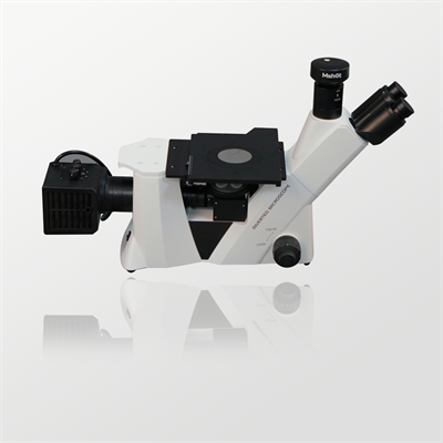
differential interference metallographic microscope
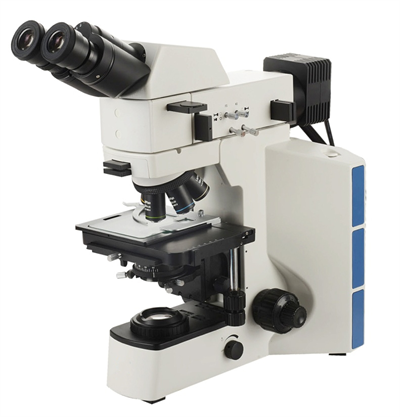
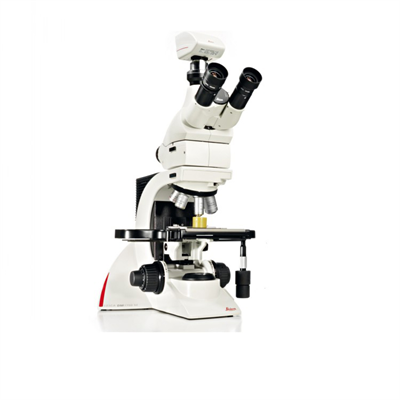
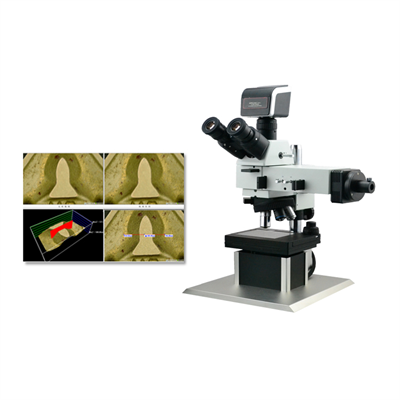
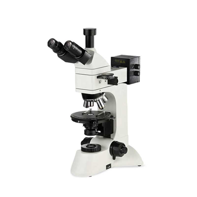
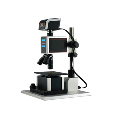
Optical Microscope
Principle: Utilizes visible light to magnify objects through a lens system, achieving high magnification through a combination of objective and ocular lenses.
Type:
Brightfield Microscope: Most commonly used, light is directly transmitted through the sample.
Dark Field Microscope: Imaging through scattered light, suitable for viewing transparent samples.
Fluorescence Microscope: Labeling samples with fluorescent dyes to excite light imaging at specific wavelengths, for dynamic observation of living cells.
Phase Difference Microscope: Enhancing contrast of transparent samples using optical path difference, suitable for viewing living cells.
Confocal Microscope: Three-dimensional imaging through laser scanning and pinhole filtering, with higher resolution.
Principle of electron microscopy: Instead of light, an electron beam is used to focus through an electromagnetic lens, with a much higher resolution than an optical microscope.
Type:
Transmission electron microscope (TEM): The electron beam penetrates the ultra-thin sample, forming an image of the internal structure.
Scanning electron microscope (SEM): The electron beam scans the surface of the sample and presents a three-dimensional topography through secondary electron imaging.
Scanning tunneling microscope (STM): Using the quantum tunneling effect to probe atomic-scale surface structures.